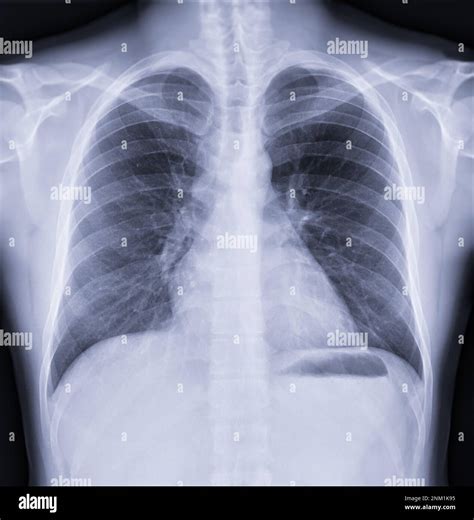
The chest X-ray is one of the most commonly ordered radiologic examinations in medicine, providing valuable information about the lungs, heart, and surrounding structures. A normal chest X-ray can be an indicator of good respiratory health, but interpreting these images requires a comprehensive understanding of what constitutes “normal” and how various factors can influence the appearance of the X-ray. Here are five key ways a normal chest X-ray can be assessed, emphasizing the importance of a thorough and nuanced interpretation:
1. Cardiothoracic Ratio

A critical aspect of assessing a chest X-ray is evaluating the cardiothoracic ratio, which is the ratio of the widest diameter of the heart to the widest diameter of the chest (from one inner edge of the rib cage to the other). Normally, this ratio should be less than 0.5. An increased cardiothoracic ratio can indicate cardiomegaly (enlargement of the heart), which might suggest various cardiac conditions. In a normal chest X-ray, the heart size should be appropriate for the patient’s body size, and this ratio can provide a quick and useful indicator of cardiac health.
2. Lung Fields and Parenchyma

The lung fields should appear clear and dark on a normal chest X-ray, indicating the absence of consolidation or other pathologies. The lung parenchyma, which includes the alveoli, bronchioles, and capillaries, should not show any signs of disease such as opacities, nodules, or cavitations. The presence of any abnormalities in the lung fields can suggest a wide range of conditions, from infections to chronic diseases like emphysema. Thus, a thorough examination of the lung fields is essential for determining the normalcy of a chest X-ray.
3. Pleural Spaces and Costophrenic Angles

The pleural spaces, which are the areas between the lungs and the chest wall, should not contain any significant amount of fluid or air (other than what is considered normal). The costophrenic angles, where the diaphragm meets the rib cage, should be sharp and well-defined. Blunting of these angles can indicate pleural effusion, a condition where excess fluid accumulates in the pleural space, which can be a sign of various diseases, including infections, malignancies, and heart failure. A normal chest X-ray will show clear, unobstructed costophrenic angles.
4. Diaphragmatic Position and Movement

The diaphragm, the major muscle used for breathing, should be at an appropriate level and have a normal contour on a chest X-ray. The position of the diaphragm can be influenced by factors such as lung volume and gravity. In a normal inspiratory chest X-ray, the diaphragm should be at or below the 10th rib anteriorly and the 6th rib posteriorly. Abnormalities in diaphragmatic position or contour can suggest conditions affecting the lungs or the diaphragm itself. Moreover, assessing the movement of the diaphragm during breathing (through fluoroscopy or video X-ray) can provide additional insights into respiratory function.
5. Bony Thorax and Soft Tissues

Finally, a normal chest X-ray should also show a normal bony thorax and soft tissues. The bony structures, including the ribs, vertebrae, and sternum, should appear intact without any fractures or lesions. The soft tissues of the chest wall, including the breasts and the muscles, should not show any signs of disease such as masses or thickening. The mediastinum, which contains the heart, trachea, esophagus, and other vital structures, should be of normal width and density, as an enlarged mediastinum can indicate serious conditions like lymphoma or aortic aneurysm.
Conclusion

Interpreting a chest X-ray as “normal” requires a detailed examination of various anatomical structures and an understanding of how different conditions can affect the appearance of these structures on the X-ray. The cardiothoracic ratio, lung fields, pleural spaces, diaphragmatic position, and the bony thorax and soft tissues all play critical roles in assessing the normalcy of a chest X-ray. Healthcare professionals must consider these factors in the context of the patient’s overall health and clinical presentation to provide an accurate interpretation and appropriate follow-up care.
What does a normal chest X-ray indicate?

+
A normal chest X-ray indicates the absence of significant abnormalities in the lungs, heart, and surrounding structures, suggesting good respiratory health. However, it’s essential to consider the X-ray in the context of the patient’s symptoms and medical history.
How is the cardiothoracic ratio calculated?

+
The cardiothoracic ratio is calculated by dividing the widest diameter of the heart by the widest diameter of the chest (from one inner edge of the rib cage to the other). A normal ratio is typically less than 0.5.
What can cause an abnormal chest X-ray?
+
A wide range of conditions can cause an abnormal chest X-ray, including infections (like pneumonia), chronic diseases (such as emphysema), heart conditions (like cardiomegaly), and malignancies. Even certain non-respiratory conditions can affect the appearance of a chest X-ray.